1. Which of the following statements about the molecular arrangement of actin in myofibrils is
incorrect?
1. Each actin (thin) filament is made of two ‘F’ (filamentous) actins helically wound to each
other.
2. Each ‘F’ actin is a polymer of monomeric ‘G’ (Globular) actins.
3. Two filaments of another protein, tropomyosin also runs close to the ‘F’ actins throughout
its length.
4. A complex protein troponin is distributed at regular intervals on the tropomyosin.
a) 1 and 2 only
b) 3 only
c) Only 4
d) none of these
Explanation: None of these
2. Select the total number of true statements from the following.
1. Each myosin (thick) filament is also a polymerized protein.
2. Many monomeric proteins called meromyosins constitute one thick filament.
3. Each meromyosin has two important parts, a globular head with a short arm and a tail,
the former being called the heavy meromyosin (HMM) and the latter is called the light
meromyosin (LMM).
4. The HMM component, i.e., the head and short arm projects outwards at regular distance
and angle from each other from the surface of a polymerized myosin filament and is
known as cross arm.
5. The globular head is an active ATPase enzyme and has binding sites for ATP and active
sites for actin.
a) 1
b) 2
c) 4
d) 5
Explanation: 5
3. Binding of Ca2+ with _____ in the skeletal muscles and leads to the exposure of the binding
site for _____ on the filament _____.
a) Troponin, myosin, actin
b) Troponin, actin, relaxin
c) Actin, myosin, troponin
d) Tropomyosin, myosis, actin
Explanation: Troponin, myosin, actin
4. Following is the figure of actin (thin) filaments. Identify A, B and C.
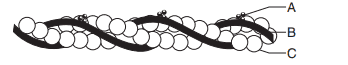
a) A−Tropomyosin, B−Troponin, C−F actin
b) A−Tropomyosin, B−Myosin, C−F Tropomyosin
c) A−Troponin, B−Tropomyosin, C−Myosin
d) A−Troponin, B−Tropomyosin, C−F actin
Explanation: A−Troponin, B−Tropomyosin, C−F actin
5.
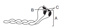
The above figure is related to the myosin monomer (meromyosin). Identify A to C.
a) A–head, B–cross arm, C–GTP binding sites
b) A−Cross arm, B−ATP Binding sites, C−Head
c) A–head, B–cross arm, C–ATP binding sites
d) A–cross arm, B–head C–ATP binding sites
Explanation: A−Cross arm, B−ATP Binding sites, C−Head
6. Identify A to D in the below figure.
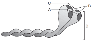
a) A–Actin binding sites, B–Head, C–Cross arm, D–ATP binding sites
b) A–Cross arm, B–Actin binding sites, C–ATP binding sites, D–Head
c) A–ATP binding sites, B–Head, C–Actin binding sites, D–Cross arm
d) A–Head, B–Cross arm, C–ATP binding sites, D–Actin binding sites
Explanation: A–ATP binding sites, B–Head, C–Actin binding sites, D–Cross arm
7. Mechanism of muscle contraction is best explained by
a) All or no law
b) Sliding filament theory
c) Blackman’s law
d) All of these
Explanation: Sliding filament theory
8. ATP provides energy for muscle contraction by allowing for
a) An action potential formation in the muscle cell.
b) Cross-bridge detachment of myosin from actin.
c) Cross-bridge attachment of myosin to actin.
d) Release of \[Ca^{2+}\] from sarcoplasmic reticulum.
Explanation: Cross-bridge attachment of myosin to actin
9. A motor unit is best described as
a) All the nerve fibres and muscle fibres in a single muscle bundle.
b) One muscle fibre and its single nerve fibre.
c) A single motor neuron and all the muscle fibres that it innervates.
d) As the neuron which carries the message from muscles to CNS.
Explanation: A motor unit is best described as a single motor neuron and all the muscle fibres that it innervates.
10. Motor end plate is a
a) Neuromuscular junction
b) Plate of motor neuron
c) Dendron of motor neuron
d) Gradient of protein motive force
Explanation: Motor end plate is a neuromuscular junction.